Blog

Your Annual Eye Exam Is Important

Berks Eye Physicians & Surgeons, Ltd.

snyderI had an interesting case the other day that does a good job of exemplifying the need for a thorough annual eye exam including dilation.

I have been seeing this patient, a 45 year old male, on a yearly basis for the last three years. It had been about 12 months so, on this visit, I did a thorough investigation of his total ocular status, including a check of his visual acuity, eye movements, field of vision, pupils, and generated an updated eyeglasses prescription as well. I then dilated his pupils after checking the health of the front part of his eyes.

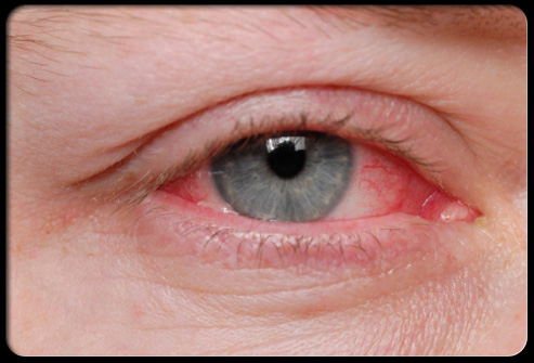
Once his pupils were large enough to adequately look through, I noticed on my direct views of the fundus (the back of the eye) several small retinal hemorrhages in his right eye. Retinal hemorrhages are areas where blood has “leaked” out of a blood vessel into the surrounding tissue. Though retinal hemorrhages can be due to a number of causes, the size, location, and appearance can tell us as eye doctors a lot of information.

Diabetes Revealed
The most common reason for new retinal hemorrhages is typically either diabetes or high blood pressure. He denied being diabetic or hypertensive but had not had a routine physical exam in over a year as well. Nonetheless, I checked his blood pressure which was normal. I then requested he make an appointment with his primary doctor for a full examination with blood work to determine the cause of the retinal hemorrhages. At his follow-up exam about a month later he reported that, indeed, he did have high blood sugar and is now being treated for Type 2 Diabetes.

Here are some interesting statistics about Diabetes:

  • In 2012, 29.1 MILLION Americans, or 9.3% of the population, had diabetes. Almost 1 in every 10 people!
  • Of those, 8.1 million were undiagnosed (like my patient).
  • In 2005–2008, of adults with diabetes aged 40 years or older, 4.2 million (28.5%) people had diabetic retinopathy, damage to the small blood vessels in the retina that may result in loss of vision.
  • Diabetes remains the 7th leading cause of death in the United States.
  • The total cost of diagnosed diabetes in the United States is estimated at $245 billion.

Of course, I don’t encounter such urgent findings on most complete eye exams. But you would be amazed how frequently I do encounter undiagnosed problems, problems that I find outright or, through conversation, a patient and I discover together – like eye allergies, contact lens comfort issues, risk factors for glaucoma or macular degeneration, glare issues from cataracts, and the ever ubiquitous DRY EYES!  It goes on and on. The point is that while problems like these probably won’t happen to you, if they do, it is much better to find out before they cause a major problem rather than react to it once they already have.

Now go out there and get those eyes checked!

By: Dr. J. Mark Snyder

Spring Into Action (before Your Allergies Do)

Berks Eye Physicians & Surgeons, Ltd.

allergies--0-24474800-1395850138-100As the winter temperatures finally start to rise, so to will the inevitable blossoms and buds marking the start of Spring. While these changes are a welcome respite from the cold, for many of us there is a bittersweet realization that seasonal allergies are coming as well.

If you don’t suffer from seasonal allergies, chances are you know at least a few people that do. In the United States, seasonal and perennial allergies affect 20% of the population, and 70% to 80% of these patients report that their allergies include red, itchy, watery eyes. 1,2

Cool compresses and avoidance of the offending allergen may suffice for mild, momentary symptoms, but they often aren’t enough or practical on a day-to-day basis. Topical relief for symptoms, however, can be very effective in relieving symptoms and restoring quality of life.

But which type of medication is the most effective? Answering this question requires an understanding of the allergic reaction in the first place. Like other allergies, allergic conjunctivitis (red, itchy eyes) is caused by a hypersensitivity reaction to something in the environment.

2 Exposure to these allergens triggers something in the body called mast cells to release histamine, among other things, that induce symptoms such as itching eyes and redness. A mast cell is like a piñata that you don’t want to open, with histamine being a very itchy type of candy. Additional symptoms include eyelid swelling and tearing. While these symptoms may mimic “pink eye” to some degree, the major difference involves the presence of itching. 3,4

A number of over-the-counter topical agents are available (such as naphazoline) – these drugs are effective at reducing redness but do very little to treat itchy eyes. 5

Having no effect on the mast cells responsible for releasing the agents that cause ocular allergy symptoms, these drugs often don’t work well enough to treat your symptoms. Another group of eye drops are called “mast cell stabilizers”. If our metaphorical piñata doesn’t open, histamine can’t be released which can prevent itching from occurring. These medications first became available for topical ophthalmic use in the 1980s.6,7

While these drugs help in reducing itchy eyes, they are relatively short-acting. To be effective they must be taken before exposure to known allergens. This strategy takes some planning and won’t be very effective for someone who is already experiencing symptoms.
A third group of drops, called antihistamines, work well for acute symptoms, especially itching. Some of the first eye drops on the market were relatively short-acting, and used to treat both redness and itchiness in the eyes.8,9  Some of these drops are still available today as non-prescription drops but they have been replaced in recent years by more effective antihistamines. A noteworthy aspect of antihistamine drops is that, unlike the pure mast cell stabilizers, these drugs are effective whenever someone experiences allergic symptoms.
A progression of antihistamines has occurred in recent years, including Asteline ®, Bepreve ®, Zaditor ® and Patanase ®. Each of these are “dual-action” antihistamines that also stabilize mast cells.10-13 Meaning, they not only prevent allergy symptoms from developing but also relieve any itching that still may occur. In addition to this dual-action effect (or perhaps because of it), these newer drugs all last longer than their predecessors. Most recently, two of them have been approved for once-daily dosing—a higher concentration formula (0.2%) of olopatadine hydrochloride, (Pataday, Alcon), and the newest ocular antihistamine, alcaftadine (Lastacaft, Allergan). 14,15

While not all patients get the appropriate relief from these medications, they are a recommended starting point in therapy given how safe they are, with limited side effects. Ultimately, knowing which drop or treatment method is best for you should be done by your eyecare professional. To take advantage of the most effective treatments for ocular allergies, time is of the essence. It is important to know your options before symptoms already are impacting your daily life. Don’t get stuck in the weeds, make an appointment to be examined today.

J. Mark Snyder, O.D.

REFERENCES:
1.     Singh K, Axelrod S, Bielory L. The epidemiology of ocular and nasal allergy in the United States, 1988-1994. J Allergy Clin Immunol. 2010 Oct;126(4):778-83.e6.
2.     Blaiss MS. Allergic rhinoconjunctivitis: burden of disease. Allergy Asthma Proc. 2007 Jul-Aug; 28(4):393-7.
3.     Akdis CA, Blaser K. Histamine in the immune regulation of allergic inflammation. J Allergy Clin Immunol. 2003 Jul;112(1):15-22.
4.     Collum LT, Kilmartin DJ. Acute allergic conjunctivitis. In: Allergic Diseases of the Eye. Philadelphia: W.B. Saunders Co.;2000:108-32.
5.     Abelson MB, Yamamoto GK, Allansmith MR. Effects of ocular decongestants. Arch Ophthalmol. 1980 May; 98(5):856-8.
6.     Abelson MB, Wun PJ, Nevius JM. Mast cell stabilizers. In: Allergic Diseases of the Eye. Philadelphia: W.B. Saunders Co.;2000:228-34.
7.     Cook EB, Stahl JL, Barney NP, Graziano FM. Mechanisms of antihistamines and mast cell stabilizers in ocular allergic inflammation. Curr Drug Targets Inflamm Allergy. 2002 Jun;1(2):167-80.
8.     Dockhorn RJ, Duckett TG. Comparison of Naphcon-A and its components (naphazoline and pheniramine) in a provocative model of allergic conjunctivitis. Curr Eye Res. 1994 May;13(5):319-24.
9.     Abelson MB, Paradis A, George MA, et al. Effects of Vasocon-A in the allergen challenge model of acute allergic conjunctivitis. Arch Ophthalmol. 1990 Apr;108(4):520-4.
10.  Giede C, Metzenauer P, Petzold U, Ellers-Lenz B. Comparison of azelastine eye drops with levocabastine eye drops in the treatment of seasonal allergic conjunctivitis. Curr Med Res Opin. 2000;16(3):153-63.
11.  Greiner JV, Mundorf T, Dubiner H, et al. Efficacy and safety of ketotifen fumarate 0.025% in the conjunctival antigen challenge model of ocular allergic conjunctivitis. Am J Ophthalmol. 2003 Dec;136(6):1097-105.
12.  Sharif NA, Xu SX, Miller ST, et al. Characterization of the ocular antiallergic and antihistaminic effects of olopatadine (AL-4943A), a novel drug for treating ocular allergic diseases. J Pharmacol Exp Ther. 1996 Sep;278(3):1252-61.
13.  Macejko TT, Bergmann MT, Williams JI, et al. Multicenter clinical evaluation of bepotastine besilate ophthalmic solutions 1.0% and 1.5% to treat allergic conjunctivitis. Am J Ophthalmol. 2010 Jul;150(1):122-27.e5.
14.  Abelson MB, Spangler DL, Epstein AB, et al. Efficacy of once-daily olopatadine 0.2% ophthalmic solution compared to twice-daily olopatadine 0.1% ophthalmic solution for the treatment of ocular itching induced by conjunctival allergen challenge. Curr Eye Res. 2007 Dec; 32(12):1017-22.
15.  Torkildsen G, Shedden A. The safety and efficacy of alcaftadine 0.25% ophthalmic solution for the prevention of itching associated with allergic conjunctivitis. Curr Med Res Opin. 2011 Mar;27(3):623-31.

Eye Allergies

Berks Eye Physicians & Surgeons, Ltd.

Eye Allergies blog of Berks EyeKeratoconus

Eye allergies are overreactions of the body’s immune system when certain substances come in contact with the eyes of sensitive individuals.

These allergy-causing substances (called allergens) can include dust, pollen, mold and
animal dander. Certain ingredients in cosmetics and eye drops also can cause eye allergies in some individuals.

Eye allergy signs and symptoms

The most common signs and symptoms of eye allergies include red eyes, swollen eyelids,
itchy eyes and excessive tearing.

Eye allergy treatment


Avoidance.The best way to handle eye allergies is to
avoid exposure to known allergens that are causing your problems. For example, if animal dander appears to be the
problem, don’t allow your pet in your bedroom and avoid touching your eyes after handling your pet. Also, purchase a
high-quality furnace filter that traps airborne pet dander.

Medications.If you’re not sure what’s causing your
eye allergies, or you’re not having any luck avoiding known allergens that are causing problems, use
doctor-recommended medication to alleviate your eye allergy symptoms.

Over-the-counter and prescription medications each have their advantages; for example,
over-the-counter products often are less expensive, while prescription ones usually are stronger and might be more
effective.

Eye drops are available as simple eye washes, or they may have one or more active
ingredients such as antihistamines, decongestants or mast cell stabilizers to relieve allergy symptoms or reduce the
severity of allergic reactions. Antihistamines relieve many symptoms caused
by airborne allergens, such as itchy, watery eyes, runny nose and sneezing.

Decongestantsclear up redness. They contain
vasoconstrictors, which make the blood vessels in your eyes smaller, lessening the apparent redness. They treat the
symptom, not the cause.

In fact, with extended use, the blood vessels can become dependent on the vasoconstrictor
to stay small. When you discontinue the eye drops, the vessels might actually get bigger than they were in the first
place. This process is called rebound hyperemia, and the result is that your red eyes worsen over time.

Some products have ingredients called mast cell stabilizers,
which alleviate redness and swelling. Mast cell stabilizers are best taken prior to allergy symptoms to reduce your
sensitivity to problematic allergens and can provide long-lasting relief.

Other medications used for eye allergies include non-steroidal anti-inflammatory drugs (NSAIDs) or corticosteroids.,
In some cases, combinations of medications may be used.

Immunotherapy.You also might benefit from
immunotherapy, in which an allergy specialist injects you with small amounts of allergens to help your body
gradually build up immunity to them.

Eye allergies and contact lenses


Even if you are a successful contact lens wearer, allergy season can make your contacts
uncomfortable. Airborne allergens can get on your lenses, causing discomfort.

Allergens also can stimulate the excessive production of natural substances in your tears
that bind to your contacts, adding to your discomfort and allergy symptoms.

Ask your eye doctor about eye drops that can help relieve your symptoms and keep your
contact lenses clean. Certain drops can discolor or damage contact lenses, so ask your doctor first before trying
out a new brand.

Another alternative is daily disposable contact lenses, which are designed to be worn once,
and then discarded at the end of the day. Because you replace them daily, these lenses are unlikely to develop
irritating deposits that can build up over time and cause or heighten allergy-related discomfort.

Article ©2011 Access Media Group LLC. All rights reserved. Reproduction other than for one-time personal use
is strictly prohibited.

Dry Eye Syndrome

Berks Eye Physicians & Surgeons, Ltd.

Keratoconus

Dry Eye Syndrome Reading PA

Dry eye syndrome (or “dry eye”) is the chronic lack of sufficient lubrication and moisture on the surface of the eye, causing discomfort, contact lens intolerance and increased risk of eye infections.

Dry eye is common, with some studies reporting that 10 to 20 million Americans experience some degree of dry eye syndrome.

Common risk factors for dry eye include increasing age, a dry environment and use of certain medications.

Signs and symptoms of dry eye

Signs and symptoms of dry eye syndrome include:

  • Red, irritated eyes
  • A burning or scratchy sensation
  • Feeling something is “in” your eye (called a foreign body sensation)
  • Fluctuating or blurred vision
  • Eye pain
  • Contact lens discomfort

It may seem odd, but another symptom of dry eye syndrome is a watery eye. This is because, as a reaction to dry eye irritation, the tear glands sometimes secrete very watery tears as a protective mechanism to prevent eye damage from a dry eye condition.

What causes dry eyes?


Dry eye is caused by the tear glands failing to secrete an adequate amount of tears or producing a tear film that, because of insufficient oiliness, evaporates too quickly.

These problems can be due to aging or a side effect of many medications, such as antihistamines, antidepressants, certain blood pressure medicines and birth control pills.

Dry eye can also be caused by chronic exposure to a dry, dusty or windy climate with low humidity. Air conditioning and forced air heating systems at home and at work also can dry out your eyes.

Another cause is failing to blink your eyes normally to remoisten them. This frequently occurs during computer work.

Dry eye syndrome also is associated with certain systemic diseases such as lupus, rheumatoid arthritis, rosacea and Sjogren’s syndrome.

Long-term contact lens wear, incomplete closure of the eyelids, eyelid disease and a deficiency of the tear-producing glands are other causes.

Dry eye syndrome is more common in women, possibly due to hormone fluctuations. Recent research suggests that smoking, too, can increase your risk of dry eye syndrome.

Dry eye also has been associated with incomplete lid closure following blepharoplasty — a popular cosmetic surgery to eliminate droopy eyelids.

Treatment for dry eye


There are several treatments for dry eye, based on the severity of the condition.

For mild dry eye, your eye doctor might recommend artificial tears, which are lubricating eye drops that are designed to alleviate the dry, scratching feeling and foreign body sensation of dry eye. Prescription eye drops for dry eye go one step further: they help increase your tear production.

If you wear contact lenses, be aware that some artificial tears and lubricating eye drops cannot be used during contact lens wear. You may need to remove your lenses before using the drops and wait 15 minutes or longer (check the label) before reinserting them.

Use only the brand of artificial tears your eye doctor recommends. Avoid self-medicating a dry eye condition by choosing artificial tears randomly in a drug store. Some products might actually make your symptoms worse.

To reduce the effects of sun, wind and dust on dry eyes, wear sunglasses when outdoors. Close-fitting wraparound styles offer the best protection.

Indoors, an air cleaner can filter out dust and other particles from the air, while a humidifier adds moisture to air that’s too dry because of air conditioning or heating.

For more significant cases of dry eye, your eye doctor might recommend punctal plugs. These tiny devices are inserted in the tear drainage ducts in your eyelids to slow the drainage of tears away from your eyes, thereby keeping your eyes more moist.

Doctors sometimes recommend special nutritional supplements such as flaxseed oil or fish oil to decrease dry eye symptoms. Drinking more water also may relieve dryness symptoms.

If medications are the cause of dry eyes, switching to a different medication or a different medical treatment may resolve the problem. However, always consult with your doctor before switching or discontinuing any medication.

Treating any underlying eyelid disease, such as blepharitis, helps as well. This may call for antibiotic or steroid drops, plus frequent eyelid scrubs with an antibacterial shampoo.

Article ©2011 Access Media Group LLC. All rights reserved. Reproduction other than for one-time personal use is strictly prohibited.

Amblyopia (lazy Eye)

Berks Eye Physicians & Surgeons, Ltd.

Amblyopia (Lazy Eye) blogKeratoconus

Amblyopia is reduced vision in an eye caused by abnormal visual development. Commonly called “lazy eye,” amblyopia usually occurs in just one eye, but both eyes can be affected.

Left untreated, amblyopia can cause legal blindness in the affected eye. About 2 to 3 percent of the American population has amblyopia.

What causes amblyopia?


The most common cause of amblyopia is strabismus, which is misalignment of the eyes. To avoid double vision caused by strabismus, the visual part of the brain suppresses visual information provided by one eye, causing that eye to be amblyopic.

Another cause of amblyopia is a significant difference in the refractive errors (nearsightedness, farsightedness and/or astigmatism) in the two eyes.

It’s important to correct amblyopia as early as possible to enable proper visual development and normal visual acuity in both eyes.

Amblyopia signs and symptoms

Amblyopia generally starts at birth or during early childhood. Because the vision in one eye usually remains normal, sometimes there are no symptoms of amblyopia until the “good” eye is covered.

If amblyopia is caused by strabismus, it is the misalignment of the eyes that often leads to the diagnosis of amblyopia after vision testing of each eye is performed.

Treatment of amblyopia


If amblyopia is caused only by unequal refractive error, sometimes full-time wear of glasses or contact lenses will be sufficient for vision to develop properly in the amblyopic eye. But in most cases, eye patching or some other technique to temporarily reduce the visual acuity of the “good” eye is needed to stimulate the visual development of the amblyopic eye.

Eye patching may be required for several hours each day or even all day long, and may continue for weeks or months. If you have a lot of trouble with your child taking the patch off, you might consider a prosthetic contact lens that is specially designed to block vision in one eye and is colored to closely match the other eye.

Another alternative to patching is the use of atropine eye drops in the non-amblyopic eye. The drops blur the vision of the good eye to force greater use of the amblyopic eye. Studies have shown use of atropine eye drops is comparable to eye patching for treating amblyopia and doesn’t require constant vigilance to make sure your child wears an eye patch.

In cases when the amblyopia is caused by a large eye turn, strabismus surgery is usually required to straighten the eyes. The surgery corrects the muscle problem that causes strabismus so the eyes can focus together and see properly.

In many cases, a program of active vision therapy also is recommended to speed the development of normal vision and visual skills in an eye with amblyopia. Vision therapy exercises the eyes and helps both eyes work as a team. Vision therapy for someone with amblyopia forces the brain to use the amblyopic eye, thus restoring vision.

Amblyopia does not go away on its own, and untreated amblyopia can lead to permanent visual problems and poor depth perception. If your child has amblyopia and his or her “good” eye develops disease or is injured later in life, this could cause a permanent disability.

For best results, amblyopia should be treated as soon as possible during childhood. If amblyopia is detected and aggressively treated before the age of 8 or 9, in many cases normal 20/20 vision can be achieved.

Article ©2011 Access Media Group LLC. All rights reserved. Reproduction other than for one-time personal use is strictly prohibited.

Keratoconus

Berks Eye Physicians & Surgeons, Ltd.

Keratoconus Treatment Reading PAKeratoconus

Keratoconus is a progressive eye disease in which degenerative changes occur in the clear
front surface of the eye (cornea), causing it to bulge outward in an irregular, cone-like shape, causing distorted
vision that cannot be corrected with glasses or soft contact lenses. It can occur in one or both eyes. Keratoconus
is relatively rare. Most studies indicate it occurs in less than 0.6 percent of the U.S. population. Onset of the
disease usually occurs in people in their teens or early twenties.

What causes keratoconus?


Research suggests the weakening of the corneal tissue that leads to keratoconus may be due
to an imbalance of enzymes within the cornea. This imbalance makes the cornea more susceptible to oxidation from
compounds called free radicals, which causes it to weaken and bulge forward. Risk factors for oxidative damage and
weakening of the cornea include a genetic predisposition, explaining why keratoconus often affects more than one
member of the same family. Keratoconus also is associated with overexposure to ultraviolet rays from the sun,
excessive eye rubbing, a history of poorly fit contact lenses and chronic eye irritation.

Signs and symptoms of keratoconus


Keratoconus usually appears in a person’s late teens or early twenties, and its onset can
be slow and gradual or relatively rapid. As the cornea becomes more irregular in shape, it causes a progressive
increase in nearsightedness and irregular astigmatism, creating problems with distorted and blurred vision. Other
symptoms include glare and light sensitivity and frequent eyeglass prescription changes.

Keratoconus treatment


In very early and mild keratoconus, vision with eyeglasses or soft contact lenses may
remain acceptable. But as the disease progresses and the cornea thins and becomes increasingly more irregular in
shape, glasses or soft contacts no longer provide adequate vision correction. Treatments for moderate and advanced
keratoconus include:

Gas permeable contact lenses: Because they are
rigid, gas permeable (GP) contacts replace the irregular shape of the central cornea of a keratoconic eye with a
smooth, uniform refracting surface for better vision correction. In some cases, large-diameter GP lenses called
scleral contact lenses may be used to vault over the entire corneal surface to correct vision problems caused by
keratoconus.

“Piggybacking” contact lenses: Because fitting a gas
permeable contact lens over a cone-shaped cornea can sometimes be uncomfortable for the individual with keratoconus,
some eye care practitioners advocate fitting the eye with a soft contact lens, and then fitting a GP contact lens
over the soft lens. With this “piggybacking” technique, the underlying soft lens acts as a cushion between the GP
lens and the corneal surface for greater comfort.

Hybrid contact lenses: Hybrid contact lenses are
premium contacts that have a GP optical center, surrounded by a “skirt” of soft lens material. These lenses are
designed to provide the optical clarity of a gas permeable contact lens, combined with wearing comfort comparable to
soft contact lenses. Special hybrid lens designs are available for the correction of keratoconus.

Intacs: These tiny plastic inserts are surgically
placed just under the eye’s surface in the periphery of the cornea, and help re-shape the cornea for clearer vision.
Intacs may be needed when keratoconus patients no longer can obtain functional vision with contact lenses or
eyeglasses.

CXL: Corneal collagen cross-linking (CXL) is a
relatively new procedure that strengthens bonds between connective tissue (collagen) fibers within the cornea to
halt the progression of keratoconus. The surface layer of the cornea (epithelium) is gently removed and the
underlying corneal tissue is treated with eye drops of riboflavin (vitamin B2) and then exposed to ultraviolet light
for several minutes. Within a few days of the brief in-office treatment, the epithelial layer grows back. Early
results of CXL research show the procedure can successfully stop the progression of keratoconus, and some patients
experience reversal of the condition and vision improvement.

Corneal transplant: In advanced cases of keratoconus
that cannot be successfully treated by other means, the last remedy is a cornea transplant, also called a
penetrating keratoplasty (PK or PKP). Even after a successful cornea transplant, most keratoconic patients still
need glasses or contact lenses for clear vision.

Article ©2011 Access Media Group LLC. All rights reserved. Reproduction other than for one-time personal use
is strictly prohibited.

Let Us Solder Your Eyeglasses

Berks Eye Physicians & Surgeons, Ltd.

Let Us Solder Your Eyeglasses at Berks Eye

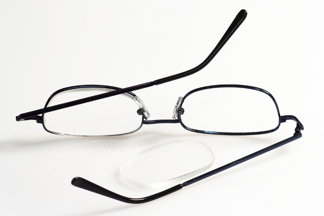
Berks Eye is unique in more ways than one – we are one of the few places that offer frame soldering right in our office. Bring in your frames and we will evaluate their condition. We have our very own laboratory with state-of-the-art equipment and lab technicians with over 20 years experience. Here, we can assure you of the highest quality workmanship in eyewear. If you have any questions at all, please feel free to call our office!

ADDRESS:
1802 Paper Mill Road Wyomissing, PA 19610
PHONE:
FAX:
(610) 376-6968
OFFICE HOURS
Monday
8:00 am - 7:00 pm
Tuesday
8:00 am - 5:00 pm
Wednesday
8:00 am - 5:00 pm
Thursday
8:00 am - 7:00 pm
Friday
8:00 am - 5:00 pm