 1802 Paper Mill Road, Wyomissing, PA 19610
What Is a Retinal Vein Occlusion (RVO)?
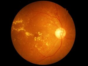
The retina is nourished through a complex network of blood vessels. Arteries transport blood to the retina, and veins drain blood away from the retina. If one of the veins partially or completely closes off, it is known as retinal vein occlusion. This can occur when the veins of the eyes are too narrow or there is a problem with the blood. A blockage of the main vein of the retina is called central retinal vein occlusion, and a blockage of one of the smaller veins is called branch retinal vein occlusion. When the veins are blocked, blood and other fluids can leak into the vitreous, causing additional problems.
Depending on the severity of the blockage, retinal vein occlusion can cause mild, intermittent visual symptoms like blurry or distorted vision — or worse, it can cause swelling in the macula. Without proper circulation, retinal cells can die and vision can be lost. New, abnormal blood vessels can form as a response to inadequate blood supply. Secondary complications like a spike in intraocular pressure are also a risk.
Occlusions and other problems with the retinal blood vessels require the knowledge of fellowship-trained specialists like Dr. Guri Bronner of Berks Eye Physicians & Surgeons. If you have been diagnosed with a retinal vein occlusion or are experiencing symptoms, please reach out to us today.
Retinal Vein Occlusion Symptoms
If you develop retinal vein occlusion, you may or may not experience visual symptoms. Signs and symptoms include a sudden loss of vision and the appearance of “floaters,” or objects that appear to float around in your field of vision.
Risk Factors for Retinal Vein Occlusion
- Diabetes
- High blood pressure
- Being overweight or obese
- Heart disease
- Pre-existing eye disease – e.g., glaucoma, macular edema
- Being prone to blood clots
Prevention of RVO
You cannot completely prevent retinal vein occlusion. However, if you have a health condition like diabetes or hypertension that is associated with a higher risk of the disease, managing that condition can help delay or prevent problems with your retinal blood vessels.
Diagnosing RVO
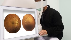
A thorough eye exam can confirm or rule out central or branch retinal vein occlusion. Our team also uses two types of retinal imaging tests when diagnosing the conditions. Fluorescein angiography involves injecting a colored dye into your arm; it travels through your veins to your eyes, and when it reaches the retinal blood vessels, we take photographs of the retina. Those photographs can help us detect blockages and other abnormalities that suggest a problem.
Optical coherence tomography, or OCT, is also useful for gathering detailed images of the retina.
Retinal Vein Occlusion Treatment Options
Treatment is always individualized to the unique needs of the patient. We will recommend the suitable treatment or combination of treatments to control leakage and swelling and restore your retina to good health.
One frequently used treatment is injections of anti-VEGF drugs, which curb swelling in the macula and prevent the growth of new, abnormal blood vessels. Laser therapy is another option to reduce the occurrence of bleeding and minimize the risk of serious complications.
Contact Us to Learn More about Retinal Vein Occlusion
For more information about central or branch retinal vein occlusion, please reach out to the team at Berks Eye Physicians & Surgeons.