 1802 Paper Mill Road, Wyomissing, PA 19610
A Glaucoma Eye Exam May Include:
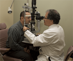 An assessment of your general health and medical history
- A review of your glaucoma risk factors
- A measurement of the visual acuity
- A measurement of the intraocular pressure (tonometry)
- A measurement of the corneal thickness (pachymetry)
- An inspection of the drainage angle of the eye (gonioscopy)
- An evaluation of the optic nerve by direct visualization for any signs of damage (ophthalmoscopy)
- An assessment of the visual field of each eye (perimetry)
Optic Nerve Imaging
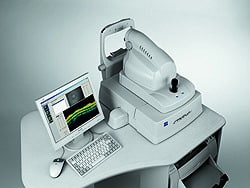
Newer, more sophisticated methods of optic nerve analysis using computerized digital imaging assist with the accuracy of the initial glaucoma diagnosis and provide a useful database of information for monitoring disease progression or stability.
Your evaluation will include Optical Coherence Tomography (OCT), which uses special light beams that reflect off the retinal surface and are captured by the computer to create images of the optic nerve or the retinal layers around the optic nerve. The measurement of the retinal nerve fiber layer thickness around the optic nerve is of particular value in a glaucoma evaluation, since loss of nerve fibers will cause thinning of the nerve fiber layer.