 1802 Paper Mill Road, Wyomissing, PA 19610
What Is Posterior Vitreous Detachment?
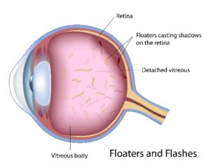
Approximately 80 percent of the volume of your eye is vitreous, a transparent substance that fills the eyeball and helps it keep its round shape. Normally the vitreous has the consistency of gel and is loosely connected to the back wall of the eye with millions of tiny collagen fibers. With advancing age, it loses its gel-like consistency and starts to liquify. The collagen fibers attaching it to the retina also deteriorate and eventually break. The vitreous no longer completely fills up the cavity and therefore starts to pull away from the retina. This separation is called posterior vitreous detachment (PVD).
Sometimes posterior vitreous detachment is harmless and causes little to no visual impairment. Occasionally the collagen fibers do not break and the vitreous can tug too hard on the retina, pulling away pieces of the light-sensitive tissue. If this occurs, it can cause complications like a retinal tear or retinal detachment. In the rare event of a retinal detachment, the eye can permanently lose vision.
If you develop posterior vitreous detachment, it should be regularly monitored by Berks Eye Physicians & Surgeons. Our team will keep a close eye on you to ensure you experience no further problems.
Posterior Vitreous Detachment Symptoms
Most cases of posterior vitreous detachment are asymptomatic. Sometimes the condition causes the illusion of tiny specks or strands (“floaters”) or flickering/flashing lights (“flashes”) in your vision. Rarely, posterior vitreous detachment can cause what appears to be a gray or dark curtain moving across your field of vision.
If you notice a sudden onset of floaters or flashes or a decline in your vision, give us a call and request an evaluation with one of our doctors.
Risk Factors of Posterior Vitreous Detachment
The biggest risk factor for posterior vitreous detachment is age. It is very rare for the condition to affect those under the age of 40. Other risk factors include the following:
- Nearsightedness
- Traumatic injury to the eye
- History of eye surgery such as cataract surgery
- Diabetes
Prevention
There is virtually no way to prevent posterior vitreous detachment. The best thing you can do for your eye health is to see our doctors for regular eye examinations.
Diagnosing Posterior Vitreous Detachment
Diagnosing posterior vitreous detachment requires an in-depth eye exam. Our team will evaluate the internal structures of your eye. Imaging technology like optical coherence tomography (OCT) is useful to detect signs of posterior vitreous detachment.
Posterior Vitreous Detachment Treatment Options
Normally treatment is only indicated if posterior vitreous detachment has led to a retinal tear or detachment. In the case of a retinal tear, we can repair the damage using laser therapy.
Contact Us Today
For more information about posterior vitreous detachment, please call or email Berks Eye Physicians & Surgeons today.