 1802 Paper Mill Road, Wyomissing, PA 19610
What Is Lattice Degeneration?
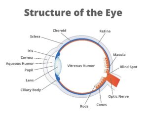
Lattice degeneration refers to an abnormal thinning of the peripheral retina. The retina is the light-sensitive tissue that lines the back wall of the eye. It converts incoming light into electrical signals that are transmitted to the brain through the optic nerve. When the retinal tissue gets abnormally thin, it is more susceptible to tears and holes. Rarely, lattice degeneration can increase the risk of retinal detachment, which is a separation of the retina from the underlying tissue. These problems can cause significant visual impairment and, if left untreated, even permanent vision loss.
The team at Berks Eye Physicians & Surgeons takes your retinal health very seriously. If you experience symptoms that suggest a problem with your retina, we encourage you to contact us promptly so we can schedule an evaluation.
Lattice Degeneration Symptoms
Lattice degeneration itself does not cause noticeable symptoms or loss of vision, but complications from the condition (e.g., retinal tear, retinal detachment) can. Signs to look out for include the following:
- Blurry vision or significant changes in visual clarity
- Frequent appearances of “flashes” – i.e., flashing or flickering lights in your visual field
- Sudden onset of “floaters” – i.e., what appears to be objects floating around in your visual field
- A dark curtain or veil that appears to obscure part of your visual field
Lattice degeneration that has not caused complications can only be detected with a dilated eye examination.
Lattice Degeneration Risk Factors
Approximately 1 in 10 people have lattice degeneration. You are more likely to get lattice degeneration if you have one or more of the following:
- Stickler syndrome, Ehlers-Danlos syndrome or Marfan syndrome
- Nearsightedness (myopia)
- A family history of lattice degeneration
Prevention
Lattice degeneration cannot be prevented.
Diagnosing Lattice Degeneration
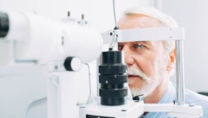
Lattice degeneration is diagnosed with a dilated eye exam. When the pupils are dilated, the doctor has a clearer view of the retina and the surrounding structures at the back of the eye. A slit lamp, or lighted microscope, is used to check for signs of lattice degeneration, including color changes, sometimes accompanied by pigment clumps and a crosshatching pattern of sclerotic (abnormal) blood vessels in the retina.
Lattice Degeneration Treatment Options
If our team detects lattice degeneration, we will regularly monitor it with dilated eye exams to ensure it does not cause any complications. We will explain what to watch for and ask you to contact us promptly if you experience any troubling symptoms. The majority of patients with lattice degeneration do not experience complications.
Sometimes we use laser therapy or cryotherapy to strengthen the thinning retina and reduce the risk of a tear or detachment.
In the event that lattice degeneration does lead to a retinal tear or detachment, we can repair it with several different types of treatment. Vitrectomy, wherein we remove the eye’s vitreous to access and repair portions of the retina, is one option. Another option is laser therapy. We will go over the pros and cons of every option and help you decide what is best for you and your vision.
Schedule a Consult with Our Team
To learn more about lattice degeneration, please call or email Berks Eye Physicians & Surgeons today.